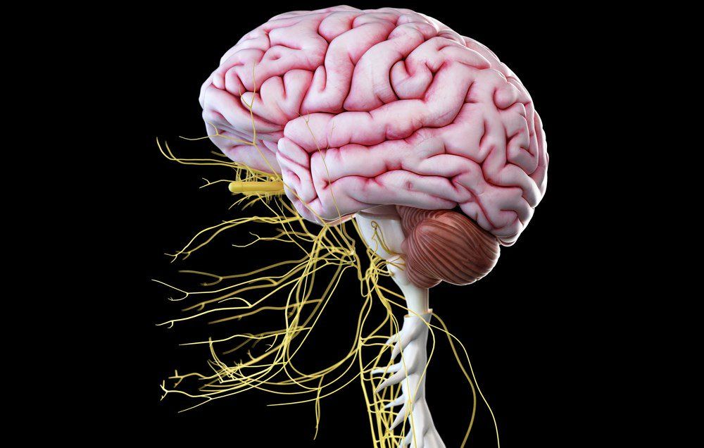
The nervous system integrates and monitors the countless actions occurring simultaneously throughout the entire human body. Therefore, every task a person accomplishes, no matter how menial, is a direct result of the components of the nervous system.
These actions can be under voluntary control, like touching a computer key, or can occur without your direct knowledge, like digesting food, releasing enzymes from the pancreas, or other unconscious acts.
It is difficult to understand all the complexities of the nervous system because the field of neuroscience has rapidly evolved over the past 20 years; moreover, answers to new questions are being found almost daily. A thorough knowledge of the individual components of the nervous system and their functions.
However, it will lead you to a better understanding of how the human body works and will facilitate your future acquisition of knowledge about the nervous system.
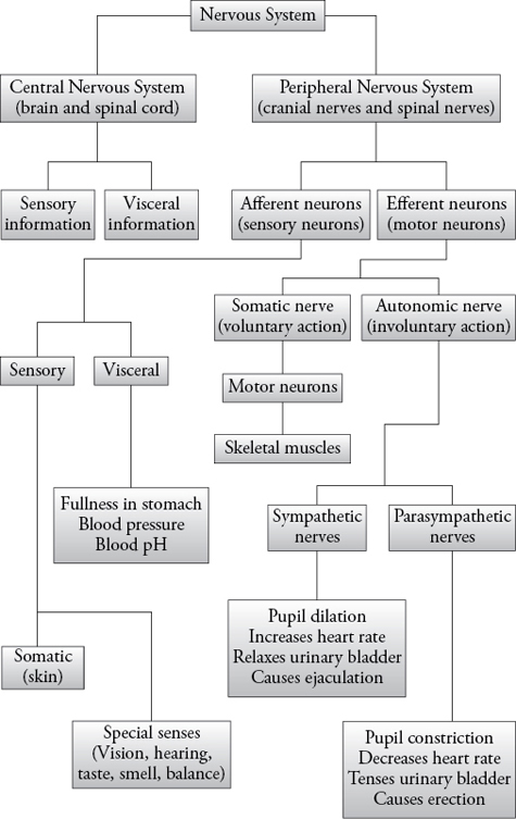
The nervous system consists of two parts;
- The central nervous system (CNS) consists of the brain and spinal cord.
- The peripheral nervous system (PNS) consists of nerves outside the CNS.
Nerves of the PNS are classified in three ways;
- First, PNS nerves are classified by how they are connected to the CNS. Cranial nerves originate from or terminate in the brain, while spinal nerves originate from or terminate at the spinal cord.
- Second, nerves of the PNS are classified by the direction of nerve propagation. Sensory ( afferent) neurons transmit impulses from skin and other sensory organs or from various places within the body to the CNS. Motor ( efferent) neurons transmit impulses from the CNS to effectors (muscles or glands).
- Third, motor neurons are further classified according to the effectors they target. The somatic nervous system (SNS) directs the contraction of skeletal muscles. The autonomic nervous system (ANS) controls the activities of organs, glands, and various involuntary muscles, such as cardiac and smooth muscles.

The Brain
Three cavities, called the primary brain vesicles, form during the early embryonic development of the brain. These are the forebrain (prosencephalon), the midbrain (mesencephalon), and the hindbrain (rhombencephalon).
During subsequent development, the three primary brain vesicles develop into five secondary brain vesicles. The names of these vesicles and the major adult structures that develop from the vesicles follow (see Table 1):
- The telencephalon generates the cerebrum (which contains the cerebral cortex, white matter, and basal ganglia).
- The diencephalon generates the thalamus, hypothalamus, and pineal gland.
- The mesencephalon generates the midbrain portion of the brainstem.
- The metencephalon generates the pons portion of the brainstem and the cerebellum.
- The myelencephalon generates the medulla oblongata portion of the brainstem.
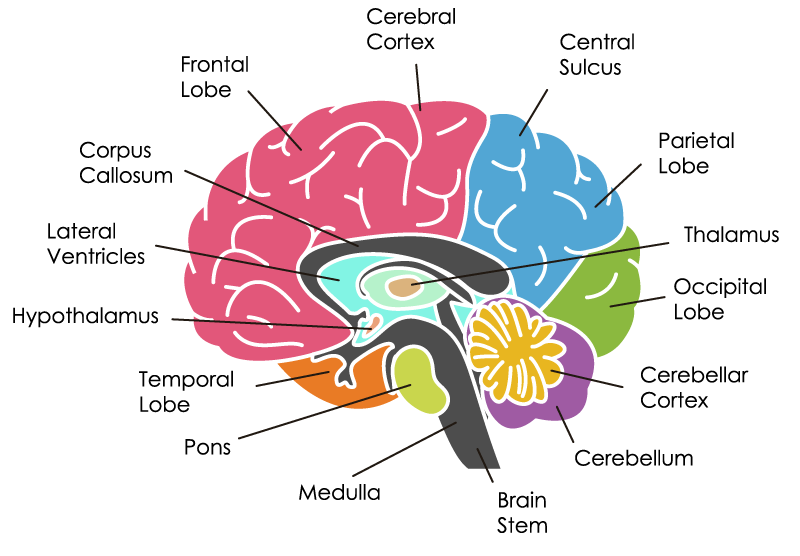
The cerebrum consists of two cerebral hemispheres connected by a bundle of nerve fibers, the corpus callosum. The largest and most visible part of the brain, the cerebrum, appears as folded ridges and grooves, called convolutions. The following terms are used to describe the convolutions:
- A gyrus (plural, gyri) is an elevated ridge.
- A sulcus (plural, sulci) is a shallow groove.
- A fissure is a deep groove.
Secrets of brain development of your child
The deeper fissures divide the cerebrum into five lobes; the frontal lobe, the parietal lobe, the temporal lobe, the occipital lobe, and the insula. All but the insula are visible from the outside surface of the brain.

A cross section of the cerebrum shows three distinct layers of nervous tissue;
- The cerebral cortex is a thin outer layer of gray matter. Such activities as speech, evaluation of stimuli, conscious thinking, and control of skeletal muscles occur here. These activities are grouped into motor areas, sensory areas, and association areas.
- The cerebral white matter underlies the cerebral cortex. It contains mostly myelinated axons that connect cerebral hemispheres (commissural fibers), connect gyri within hemispheres (association fibers), or connect the cerebrum to the spinal cord (projection fibers). The corpus callosum is a major assemblage of commissural fibers that forms a nerve tract that connects the two cerebral hemispheres.
- Basal ganglia (basal nuclei) are several pockets of gray matter located deep inside the cerebral white matter. The major regions in the basal ganglia—the caudate nuclei, the putamen, and the globus pallidus—are involved in relaying and modifying nerve impulses passing from the cerebral cortex to the spinal cord. Arm swinging while walking, for example, is controlled here.
- The diencephalon connects the cerebrum to the brainstem. It consists of the following major regions:
- The thalamus is a relay station for sensory nerve impulses traveling from the spinal cord to the cerebrum. Some nerve impulses are sorted and grouped here before being transmitted to the cerebrum. Certain sensations, such as pain, pressure, and sensitivity to temperature, are also evaluated here.
- The epithalamus contains the pineal gland. The pineal gland secretes melatonin, a hormone that helps regulate the biological clock (sleep‐wake cycles).
- The hypothalamus regulates numerous important body activities. It controls the autonomic nervous system and regulates emotion, behavior, hunger, thirst, body temperature, and the biological clock. It also produces two hormones (antidiuretic hormone or ADH, and oxytocin) and various releasing hormones that control hormone production in the anterior pituitary gland.

- The mammillary bodies relay information related to eating, such as chewing and swallowing.
- The infundibulum connects the pituitary gland to the hypothalamus.
- The optic chiasma passes between the hypothalamus and the pituitary gland. Here, portions of the optic nerve from each eye cross over to the cerebral hemisphere on the opposite side.
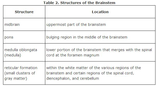
- The brainstem connects the diencephalon to the spinal cord. The brainstem resembles the spinal cord in that both consist of white matter fiber tracts surrounding a core of gray matter. The brainstem consists of the following four regions, all of which provide connections between various parts of the brain and between the brain and the spinal cord.

The reticular activation system (RAS), one component of the reticular formation, is responsible for maintaining wakefulness and alertness and for filtering out unimportant sensory information. Other components of the reticular formation are responsible for maintaining muscle tone and regulating visceral motor muscles.
The cerebellum consists of a central region (the vermis) and two winglike lobes (the cerebellar hemispheres). Like that of the cerebrum, the surface of the cerebellum is convoluted, but the gyri, called folia, are parallel and give a pleated appearance.
The cerebellum evaluates and coordinates motor movements by comparing actual skeletal movements to the movement that was intended.
The Ventricles and Cerebrospinal Fluid
There are four cavities in the brain, called ventricles. The ventricles are filled with cerebrospinal fluid (CSF), which provides the following functions:
- Absorbs physical shocks to the brain
- Distributes nutritive materials to and removes wastes from nervous tissue
- Provides a chemically stable environment
There are four ventricles:
- Each of two lateral ventricles (ventricles 1 and 2) occupies a cerebral hemisphere.
- The third ventricle is connected by a passage (interventricular foramen) to each of the two lateral ventricles.
- The fourth ventricle connects to the third ventricle (via the cerebral aqueduct) and to the central canal of the spinal cord (a narrow, central tube extending the length of the spinal cord). Additional openings in the fourth ventricle allow CSF to flow into the subarachnoid space.
A network of capillaries called the choroid plexus projects into each ventricle. Ependymal cells (a type of neuroglial cell) surround these capillaries. Blood plasma entering the ependymal cells from the capillaries is filtered as it passes into the ventricle, forming CSF.
Any material passing from the capillaries to the ventricles of the brain must do so through the ependymal cells because tight junctions linking these cells prevent the passage of plasma between them. Thus, the ependymal cells maintain a blood‐CSF barrier, controlling the composition of the CSF.
The CSF circulates from the lateral ventricles (where most of the CSF is produced) to the third and then fourth ventricles. From the fourth ventricle, most of the CSF passes into the subarachnoid space, a space within the linings (meninges) of the brain, although some CSF also passes into the central canal of the spinal cord.
The CSF returns to the blood through the arachnoid villi located in the dural sinuses of the meninges.
The Meninges
The meninges (singular, meninx) are protective coverings of the brain (cranial meninges) and spinal cord (spinal meninges). They consist of three layers of membranous connective tissue:
- The dura mater is the tough outer layer lying just inside the skull and vertebrae. Some characteristics follow:
- In the brain, there are channels within the dura mater, the dural sinuses, which contain venous blood returning from the brain to the jugular veins.
- In the spinal cord, the dura mater is often referred to as the dural sheath. A fat‐filled space between the dura mater and the vertebrae, the epidural space, acts as a protective cushion to the spinal cord.
- The arachnoid (arachnoid mater) is the middle meninx. Projections from the arachnoid, called arachnoid villi, protrude through one layer of the dura mater into the dural sinuses. The arachnoid villi transport the CSF from the subarachnoid space to the dural sinuses. Two cavities border the arachnoid:
- The subdural space occurs outside the arachnoid (between the arachnoid and the dura mater).
- The subarachnoid space lies inside the arachnoid. This space contains blood vessels and circulates CSF. The fine threads of tissue that spread across this space resemble a spider web and give the arachnoid layer its name (arachnid means spider).
- The pia mater is the innermost meninx layer. It tightly covers the brain (following its convolutions) and spinal cord and carries blood vessels that provide nourishment to these nervous tissues.
The Blood-Brain Barrier
Cells in the brain require a very stable environment to ensure controlled and selective stimulation of neurons. As a result, only certain materials are allowed to pass from blood vessels to the brain. Substances such as O2, glucose, H2O, CO2, essential amino acids, and most lipid‐soluble substances enter the brain readily.
blood‐brain barrier is established by the following:
- Brain capillaries are less permeable than other capillaries because of tight junctions between the endothelial cells in the capillary walls.
- The basal lamina (secreted by the endothelial cells) that surrounds the brain capillaries decreases capillary permeability. This layer is usually absent in capillaries found elsewhere.
- Processes from astrocytes (a type of neuroglial cell) cover brain capillaries and are believed to influence capillary permeability in some way.
Cranial Nerves
Cranial nerves are nerves of the PNS that originate from or terminate in the brain. There are 12 pairs of cranial nerves, all of which pass through foramina of the skull. Some cranial nerves are sensory nerves (containing only sensory fibers), some are motor nerves (containing only motor fibers), and some are mixed nerves (containing a combination of sensory and motor nerves).
Characteristics of the cranial nerves, which are numbered from anterior to posterior as they attach to the brain.

The Spinal Cord
The spinal cord has two functions:
- Transmission of nerve impulses. Neurons in the white matter of the spinal cord transmit sensory signals from peripheral regions to the brain and transmit motor signals from the brain to peripheral regions.
- Spinal reflexes. Neurons in the gray matter of the spinal cord integrate incoming sensory information and respond with motor impulses that control muscles (skeletal, smooth, or cardiac) or glands.
The spinal cord is an extension of the brainstem that begins at the foramen magnum and continues down through the vertebral canal to the first lumbar vertebra (L 1). Here, the spinal cord comes to a tapering point, the conus medullaris.
The spinal cord is held in position at its inferior end by the filum terminale, an extension of the pia mater that attaches to the coccyx. Along its length, the spinal cord is held within the vertebral canal by denticulate ligaments, lateral extensions of the pia mater that attach to the dural sheath.
The following are external features of the spinal cord;
- Spinal nerves emerge in pairs, one from each side of the spinal cord along its length.
- The cervical nerves form a plexus (a complex interwoven network of nerves—nerves converge and branch).
- The cervical enlargement is a widening in the upper part of the spinal cord (C 4–T 1). Nerves that extend into the upper limbs originate or terminate here.
- The lumbar enlargement is a widening in the lower part of the spinal cord (T 9–T 12). Nerves that extend into the lower limbs originate or terminate here.
- The anterior median fissure and the posterior median sulcus are two grooves that run the length of the spinal cord on its anterior and posterior surfaces, respectively.
- The cauda equina are nerves that attach to the end of the spinal cord and continue to run downward before turning laterally to other parts of the body.
- There are four plexus groups: cervical, brachial, lumbar, and sacral. The thoracic nerves do not form a plexus.

A cross section of the spinal cord reveals the following features;
- Roots are branches of the spinal nerve that connect to the spinal cord. Two major roots form the following:
- A ventral root (anterior or motor root) is the branch of the nerve that enters the ventral side of the spinal cord. Ventral roots contain motor nerve axons, transmitting nerve impulses from the spinal cord to skeletal muscles.
- A dorsal root (posterior or sensory root) is the branch of a nerve that enters the dorsal side of the spinal cord. Dorsal roots contain sensory nerve fibers, transmitting nerve impulses from peripheral regions to the spinal cord.
- A dorsal root ganglion is a cluster of cell bodies of a sensory nerve. It is located on the dorsal root.
- Gray matter appears in the center of the spinal cord in the form of the letter H (or a pair of butterfly wings) when viewed in cross section:
- The gray commissure is the crossbar of the H.
- The anterior (ventral) horns are gray matter areas at the front of each side of the H. Cell bodies of motor neurons that stimulate skeletal muscles are located here.
- The posterior (dorsal) horns are gray matter areas at the rear of each side of the H. These horns contain mostly interneurons that synapse with sensory neurons.
- The lateral horns are small projections of gray matter at the sides of H. These horns are present only in the thoracic and lumbar regions of the spinal cord. They contain cell bodies of motor neurons in the sympathetic branch of the autonomic nervous system.
- The central canal is a small hole in the center of the H crossbar. It contains CSF and runs the length of the spinal cord and connects with the fourth ventricle of the brain.
- White columns (funiculi) refer to six areas of the white matter, three on each side of the H. They are the anterior (ventral) columns, the posterior (dorsal) columns, and the lateral columns.
- Fasciculi are bundles of nerve tracts within white columns containing neurons with common functions or destinations:
- Ascending (sensory) tracts transmit sensory information from various parts of the body to the brain.
- Descending (motor) tracts transmit nerve impulses from the brain to muscles and glands.
Spinal Nerves
There are 31 pairs of spinal nerves (62 total). The following discussion traces a spinal nerve as it emerges from the spinal column:
A spinal nerve emerges at two points from the spinal cord, the ventral and dorsal roots.
- The ventral and dorsal roots merge to form the whole spinal nerve.
- The spinal nerve emerges from the spinal column through an opening (intervertebral foramen) between adjacent vertebrae. This is true for all spinal nerves except for the first spinal nerve (pair), which emerges between the occipital bone and the atlas (the first vertebra).
- Outside the vertebral column, the nerve divides into the following branches:
- The dorsal ramus contains nerves that serve the dorsal portions of the trunk.
- The ventral ramus contains nerves that serve the remaining ventral parts of the trunk and the upper and lower limbs.
- The meningeal branch reenters the vertebral column and serves the meninges and blood vessels within.
- The rami communicantes contain autonomic nerves that serve visceral functions.
- Some ventral rami merge with adjacent ventral rami to form a plexus, a network of interconnecting nerves. Nerves emerging from a plexus contain fibers from various spinal nerves, which are then carried together to some target location.
An area of the skin that receives sensory stimuli that pass through a single spinal nerve is called a dermatome. Dermatomes are illustrated on a human figure with lines that mark the boundaries of the area where each spinal nerve receives stimuli.
Reflexes
A reflex is a rapid, involuntary response to a stimulus. A reflex arc is the pathway traveled by the nerve impulses during a reflex. Most reflexes are spinal reflexes with pathways that traverse only the spinal cord.
During a spinal reflex, information may be transmitted to the brain, but it is the spinal cord, not the brain, that is responsible for the integration of sensory information and a response transmitted to motor neurons.
Some reflexes are cranial reflexes with pathways through cranial nerves and the brainstem.
- The receptor is the part of the neuron (usually a dendrite) that detects a stimulus.
- The sensory neuron transmits the impulse to the spinal cord.
- The integration center involves one synapse (monosynaptic reflex arc) or two or more synapses (polysynaptic reflex arc) in the gray matter of the spinal cord. In polysynaptic reflex arcs, one or more interneurons in the gray matter constitute the integration center.
- A motor neuron transmits a nerve impulse from the spinal cord to a peripheral region.
- An effector is a muscle or gland that receives the impulse from the motor neuron. In somatic reflexes, the effector is skeletal muscle. In autonomic (visceral) reflexes, the effector is smooth or cardiac muscle, or a gland.
The Autonomic Nervous System
The peripheral nervous system consists of the somatic nervous system (SNS) and the autonomic nervous system (ANS). The SNS consists of motor neurons that stimulate skeletal muscles.
In contrast, the ANS consists of motor neurons that control smooth muscles, cardiac muscles, and glands. In addition, the ANS monitors visceral organs and blood vessels with sensory neurons, which provide input information for the CNS.
The ANS is further divided into the sympathetic nervous system and the parasympathetic nervous system. Both of these systems can stimulate and inhibit effectors.
However, the two systems work in opposition—where one system stimulates an organ, the other inhibits. Working in this fashion, each system prepares the body for a different kind of situation, as follows:
- The sympathetic nervous system prepares the body for situations requiring alertness or strength, or situations that arouse fear, anger, excitement, or embarrassment (“fight‐or‐flight” situations). In these kinds of situations, the sympathetic nervous system stimulates cardiac muscles to increase the heart rate, causes dilation of the bronchioles of the lungs (increasing oxygen intake), and causes dilation of blood vessels that supply the heart and skeletal muscles (increasing blood supply). The adrenal medulla is stimulated to release epinephrine (adrenalin) and norepinephrine (noradrenalin), which in turn increases the metabolic rate of cells and stimulates the liver to release glucose into the blood. Sweat glands are stimulated to produce sweat. In addition, the sympathetic nervous system reduces the activity of various “tranquil” body functions, such as digestion and kidney functioning.
- The parasympathetic nervous system is active during periods of digestion and rest. It stimulates the production of digestive enzymes and stimulates the processes of digestion, urination, and defecation. It reduces blood pressure and heart and respiratory rates and conserves energy through relaxation and rest.
In the SNS, a single motor neuron connects the CNS to its target skeletal muscle. In the ANS, the connection between the CNS and its effector consists of two neurons—the preganglionic neuron and the postganglionic neuron. The synapse between these two neurons lies outside the CNS, in an autonomic ganglion.
The axon (preganglionic axon) of a preganglionic neuron enters the ganglion and forms a synapse with the dendrites of the postganglionic neuron. The axon of the postganglionic neuron emerges from the ganglion and travels to the target organ.
Read more about your digestive system
The above information is Sourced.
