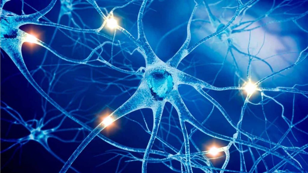
Bioelectricity is the electric potentials and currents produced by or occurring within living organisms. It is the generation or action of current or voltages in biological processes. For diagnostic purposes, the measurement of bioelectric signals has become vital in monitoring electrical effects of active cells of heart and brain. Modulating the brains bioelectricity in developing embryo could address injuries or birth defects.
In a developing embryo, the outer membrane of each cell contains protein channels. They transport negative and positive ions, producing voltage gradients across the cell wall. Groups of cells create patterns of membrane voltage. It precedes and controls the expression of genes and the morphological changes occurring over the course of development.
For the first time, Tufts University biologists have demonstrated that electrical patterns in the developing embryo can be predicted, mapped, and manipulated to inhibit deformities caused by harmful substances such as nicotine. Also targeting bioelectric states may be a new treatment method for regenerative repair in brain development and disease. This computational method can be utilized to investigate effective repair strategies.
In work published in Journal of Neuroscience, Michael Levin (Director of the Allen Discovery Center at Tufts University) and his colleagues show that brain development in frog embryos is shaped by changes in the voltage gradients across cell membranes. Thus by modulating the bioelectric signals, the researchers were able to stimulate the growth of extra brain tissue. It also helps to counterbalance the brain defects caused by a genetic mutation.
The study was more focused on gene expression, growth factors, and molecular pathways of cells. Moreover, it was unclear that how cells arrange themselves into complex organ systems in a growing embryo. Once it gets decoded, then it will help to normalize development or support regeneration in the treatment of disease or injury.
BioElectric Tissue Simulation Engine Model
Nicotine is a neuroteratogen, induces serious defects in brain patterning and learning. It causes depolarization of cells and induce positive charge. This causes disruption of normal bioelectric pattern. Further leads to birth defcets.
Hyperpolarization is a change in a cell’s membrane potential that makes it more negative (Endogenous negative charge of the cell).
One reagent, in particular, the hyperpolarization-activated cyclic nucleotide-gated channel (HCN2) rescues brain defects by enforcing endogenous bioelectric patterns, brain morphology, markers of gene expression and near normal learning capacity in the grown tadpole.
Bioelectrical signaling interrelate cell behaviors toward correct anatomical results. But uncertain information on bioelectric states makes it difficult to understand the etiology of some birth defects and the development of predictive interventions.
Michael Levin and Vaibhav Pai from Allen Discovery center at Tufts explored another computational simulation platform called BioElectric Tissue Simulation Engine (BETSE). This unique model helps to create a dynamic map of voltage signals in developing embryo of the frog.
They used the model to recognize the reagents that restore the normal pattern of bioelectric signals even in the presence of molecules that cause birth defects. In nicotine exposed embryos, the reagents helped to enhance hyperpolarization when added to the cells in the model. Thus effectively restored the normal electrical patterns.
As a matter of fact, modulating the bioelectrical signals has guided us to a different approach to diseases like cancer, birth defects, tissue regeneration and to some extent chronic diseases like diabetes. Specifically, this is an important step providing a realistic model that bridges the molecular, cellular, bioelectrical, and anatomical scales of the developing embryo.
